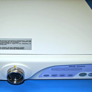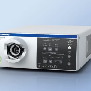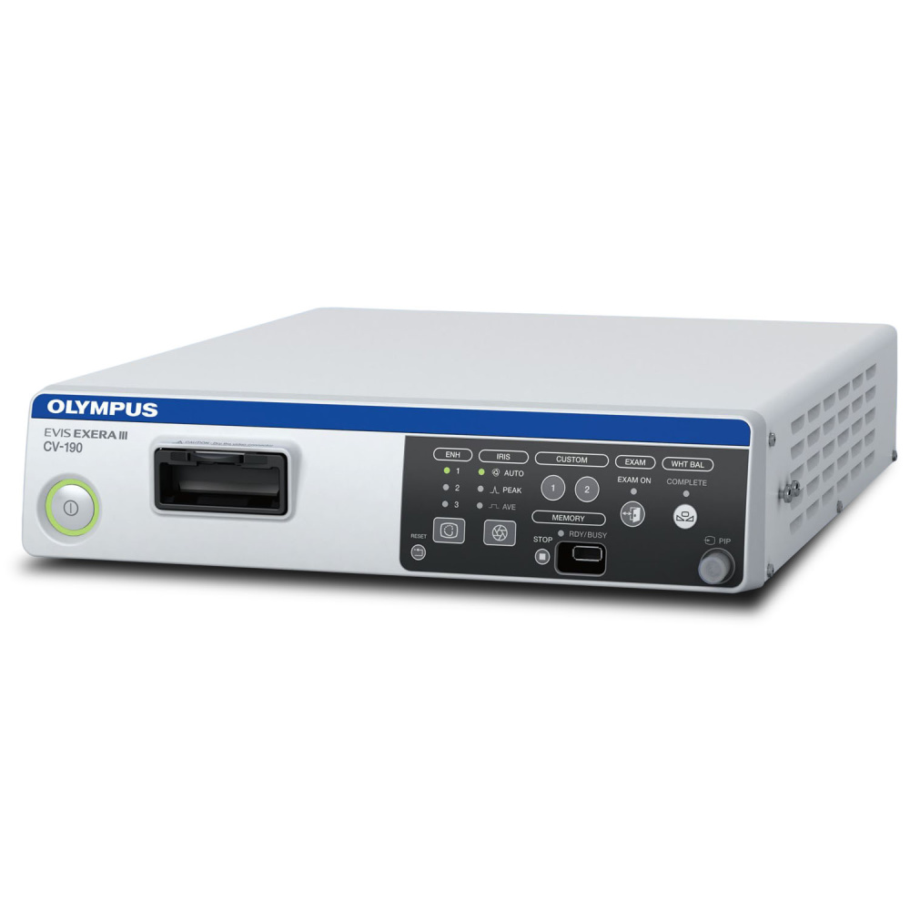
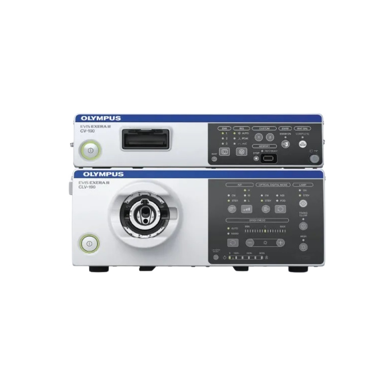
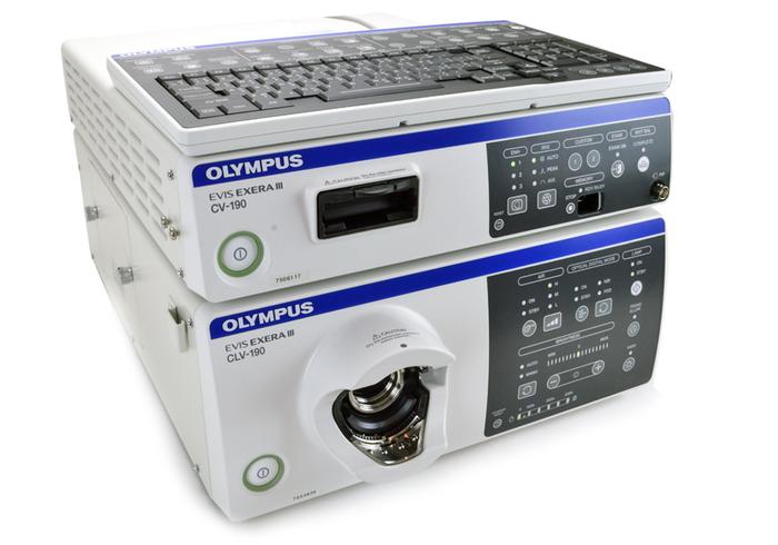
Olympus EXERA II CV-190 HDTV Compatible Video Processor. Certified pre-owned with an 18 month warranty.
The EVIS EXERA III CV-190 Video Processor provides advanced video processing capabilities. This system enables full access to the range of advanced features of the EVIS EXERA III endoscopes. New as well as improved image processing helps to deliver outstanding image quality with features such as reduced noise and improved color contrast to give the best image possible.
Key Features:
- NBI: (Narrow Band Imaging) in EVIS EXERA III 190 Series
scopes provides twice the viewable distance of EVIS
EXERA II 180 Series scopes and offers significantly brighter
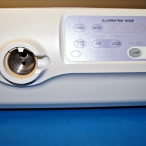
images. - Dual Focus: CV-190 features the ability to switch the point of focus
between ‘near’ and ‘normal’ with the push of a button. - Waterproof One-Touch Connector: The newly designed, waterproof one-touch connector
enables a one-step connection to the light source and does
not require a separate scope cable for the video processor. - Improved Image Processing: New and improved image processing delivers sophisticated
image quality via enhanced color reproduction, minimized
image noise, and reduced halation. - Pre-Freeze: The pre-freeze function selects the clearest still image
automatically, saving you precious time. - Full Compatibility: The CV-190 is compatible with EVIS 100/130/140/150 Series,
EVIS EXERA 160 Series, EVIS EVERA II 180 Series, GI/BF/
VISERA Series scopes, and EVIS EXERA III 190 Series. - Multiple Video Outputs Available: 16:9 and 16:10 outputs for a HDTV monitor are available.
The system is compatible with analog, HD-SDI, and DVI output. - Improved Connectivity: A link connection to peripheral devices avoids complicated
cable connections and accelerates transmission speed. - PIP: Picture-in-picture and index functions effectively enhance
your observation. - Portable Storage: The system is compatible with portable memory, which is
standard for data management; simply connect and upload.
Power Supply: Voltage 100-240 V AC (NTSC)/220-240 V AC (PAL); within ±10%
Frequency: 50/60 Hz; within ±1 Hz
Consumption electric power: 150 VA
Dimensions: (W x H x D) 370 x 85 x 455 mm / 15″ x 3.4″ x 18″
Weight: 10.7 kg / 23 lbs
Analog HDTV signal output: Either RGB (1080/60I: NTSC)/(1080/50I: PAL) or YPbPr (1080/60I: NTSC)/(1080/50I: PAL) output can be selected.
Analog SDTV signal output: VBS composite (480/60I: NTSC)/(576/50I: PAL), Y/C (480/60I: NTSC)/(576/50I: PAL), and RGB (480/60I: NTSC)/(576/50I: PAL); simultaneous outputs possible
Digital signal output: HD-SDI (SMTPE 292M), SD-SDI (SMPTE 259M), DV (IEEE 1394), and DVI (WUXGA, 1080p or SXGA) can be selected.
White balance adjustment: White balance adjustment is possible using the white balance button on the front panel.
Standard color chart output: The “Color bar” or the “50% white” screen can be displayed.
Color tone adjustment: The following color tone adjustments are possible using the color-tone-level adjustment button and color-tone selector button on the keyboard:
• Red adjustment: ±8 steps • Blue adjustment: ±8 steps • Chroma adjustment: ±8 steps
Automatic gain control (AGC): The image can be electronically amplified when the light is inadequate due to the distal end of the endoscope being too far from the object.
Contrast: • N (Normal): Normal image • H (High): The dark areas are darker and the bright areas are brighter than in the normal image.
• L (Low): The dark areas are brighter and bright areas are darker than in the normal image.
Iris: The auto iris modes can be selected using the “iris mode” switch on the front panel.
• Auto: The brightness is adjusted based on the brightest part of the central part and the average brightness of the periphery part.
• Peak: The brightness is adjusted based on the brightest part of the endoscopic image.
• Average: The brightness is adjusted based on the average brightness of the endoscopic image.
Image enhancement setting: Fine patterns or edges in the endoscopic images can be enhanced electronically to increase the image sharpness.
Either the structural enhancement or edge enhancement can be selected according to the user setup.
• Structural enhancement: Enhancement of contrast of the fine patterns in the image • Edge enhancement: Enhancement of edges of the endoscopic image
Switching the enhancement modes: The enhancement level can be selected from 4 levels (off, 1, 2, and 3) using the image enhancement mode button on the front panel.
Image size selection The size of the endoscopic image can be changed using the “IMAGE SIZE” key on the keyboard.
Freeze An endoscopic image is frozen using a “FREEZE” key on the endoscope or on the system keyboard.
Switching the method of freezing the endoscopic image Pre-freezing: The image with the least blur is selected and displayed from the images captured in the set time period before the freeze operation.
Fog-free function: When a compatible endoscope is connected to the video system center, the fog-free function can be used.
Endoscope’s remote switches function: The functions of the remote switches on the endoscope can be set in the user settings.
Reset to defaults: The following settings can be reset to their defaults using the reset button on the front panel:
• Color tone • Iris mode • Image-enhancement mode • Color-enhancement mode • Optical-digital observation • Image size • Contrast • Freeze
• Release index • Electronic zoom • Arrow pointer • Stopwatch • Characters on screen • PIP/POP
Remote control: The following ancillary equipment can be controlled (specified models only):
• Monitor • DVR • Video printer • Image filing system
Patient data: The following data can be displayed on the monitor using the keyboard:
• Patient ID • Patient name • Sex • Age • Date of birth • Date of recording (time, stopwatch) • Comments
Displaying the record state: The recording state of the following ancillary equipment can be displayed on the monitor:
• Portable memory and internal buffer • DVR • Video printer • Image filing system
Displaying the image information: The following data can be displayed on the monitor:
• Structure-enhancement level • Edge-enhancement level • Zoom ratio • Color mode • Focus
Advance registration of patient data Data: for up to 50 patients can be registered, such as:
• Patient ID • Patient name • Sex and age • Date of birth
Media: MAJ-1925 (OLYMPUS)
Recording format: • TIFF: no compression • JPEG (1/5): approx. 1/5 compression • JPEG (1/10): approx. 1/10 compression
Number of recorded images: • TIFF: approx. 227 images • JPEG (1/5): approx. 1024 images • JPEG (1/10): approx. 2048 images
User settings: Up to 20 user settings can be registered.
Memorization of selected setting: The following settings are held in memory even after the video system center is turned off:
• Color tone • Iris mode • Enhancement • Color-enhancement mode • Contrast • AGC • Color mode • White balance
Lithium battery Life: 5 years